肺气肿在临床上是属于常见病、多发病,特别是对于有肺部基础疾病、长期吸烟的人群来说,肺气肿的发病率是比较高的。常见的临床症状以呼吸困难为主,伴有咳嗽,咳痰,发热,全身乏力,心慌,体力减退等。
临床上对于肺气肿的治疗主要是以对症治疗为主。因为不管是轻度肺气肿还是重度肺气肿是很难达到根治的,根据患者出现的临床症状来对症治疗,尽可能的缓解患者的临床症状。目前选用的治疗方法主要以药物治疗和支持治疗为主,药物治疗的选择主要是引用支气管舒张药物、支气管扩张药物、激素等,改善患者支气管痉挛引起的呼吸困难。同时需要以支持治疗为辅助,给予吸氧,加强营养,高蛋白、高热量饮食等辅助治疗,有利于患者病情的恢复。同时完善相关的辅助检查肺部ct肺功能检测来评估患者病情的严重程度,以便于后续远外继续给予辅助手段治疗。
总而言之,肺气肿的治疗主要是根据患者出现的临床症状以及肺功能的检查情况来进行全面的评估,选择合适的治疗手段来尽可能的缓解患者的临床症状。在日常生活中,除了需要积极的治疗以外,尽早的戒烟、治疗肺部的基础疾病、脱离粉尘环境等都有利于肺气肿的恢复。做好相关的预防手段来防止肺气肿的复发,可以有效降低肺气肿发病率,提高生活质量。

肺气肿在呼吸内科是属于常见疾病,主要的发病原因有慢性支气管炎发展,空气污染,长期吸烟,吸二手烟,长期接触粉尘,遗传因素,自身酶的缺乏等。一般可见的症状主要是呼吸困难,憋喘,咳嗽,咳痰等,需要与肺部其他疾病进行鉴别。
早期肺气肿患者没有明显的临床症状,不需要经过任何治疗,目前临床上也没有什么特效药可以治疗早期肺气肿。对于早期肺气肿患者最好的治疗是避免不良因素来防止肺气肿的进一步加重。对于早期肺气肿患者来说要积极的戒烟,脱离粉尘环境,控制肺部慢性炎症,只有从根本上改变不良因素的刺激,才有可能防止肺气肿的进一步发展。目前临床上对于肺气肿的诊断主要是根据影像学检查以及肺功能的检查来进行诊断。
早期肺气肿患者可以没有明显的临床症状,一般是通过定期体检或者其他检查无意中确诊的,要根据患者的病情来全面评估选择合适的预防方案,才能有效的控制病情,防止病情进一步发展。虽然肺气肿在临床上并不能治愈,但是对于早期肺气肿患者,经过积极的预防手段,可以有效的延缓病情的进展。所以说,早期肺气肿患者不需要经过任何药物治疗,从根本上改变自身不良的生活饮食习惯,脱离不良的工作环境等,可以进一步减轻患者的临床症状,从而控制肺气肿的发展。

从中医角度讲,肺气肿可以分为痰浊壅肺证,痰热郁肺证和肺肾气虚证三种证型。下面咱们就一块来梳理一下吧。
痰浊壅肺型
对于痰浊壅肺型的肺气肿患者,咱们中药调理以祛痰化浊,降气平喘为治疗法则。方子可以用二陈汤合三子养亲汤加减。中药包括:苏子,郁金,白芥子,石菖蒲,莱菔子,陈皮,青皮,半夏,鱼腥草,茯苓,炙甘草,地龙,桑白皮等药一块应用。
痰热郁肺型
对于痰热郁肺型的肺气肿患者,咱们中药调理以清热化痰,降气平喘为治疗大法。方子可以用涤痰汤加减。中药包括:胆南星,陈皮,浙贝母,半夏,桑白皮,川贝母,生石膏,苦杏仁,枇杷叶,炙甘草,桔梗等药一块应用。
肺肾气虚型
对于肺肾气虚型的肺气肿患者,咱们中药调理以温补肺肾之气为治疗原则,方子可以用金匮肾气丸加减。中药包括:制附子,大枣,肉桂,生姜乌梅,生地黄,五味子,炒山药,麦冬,酒萸肉,半夏,炙甘草等药一块应用。
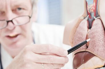
总是咳嗽咳痰、呼吸困难,肺气肿该怎样治疗?
中药治疗
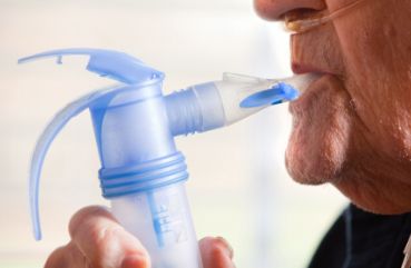
总是咳嗽咳痰、呼吸困难,肺气肿的治疗方式有哪些?
肺气肿是终末细支气管远端的气道弹性减退,过度膨胀、充气和肺容积增大或同时伴有气道壁破坏的病理状态。按其发病原因可分为老年性肺气肿、代偿性肺气肿、间质性肺气肿、灶性肺气肿、旁间隔性肺气肿、阻塞性肺气肿。
不管是哪种类型的肺气肿,都属于需要长期治疗的慢性疾病,患者一定要坚持长期治疗,切忌三天打鱼两天晒网。对于肺气肿,专家表示,在急性加重期可以使用西药来缓解症状,在缓解稳定期可使用中西结合的方式进行双管齐下的治疗。我们一起来看一下肺气肿的治疗方式有哪些?
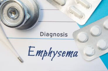
肺气肿是终末细支气管远端的气道弹性减退,过度膨胀、充气和肺容积增大或同时伴有气道壁破坏的病理状态。按其发病原因可分为老年性肺气肿、代偿性肺气肿、间质性肺气肿、灶性肺气肿、旁间隔性肺气肿、阻塞性肺气肿。
不管是哪种类型的肺气肿,都属于需要长期治疗的慢性疾病,患者一定要坚持长期治疗,切忌三天打鱼两天晒网。对于肺气肿,专家表示,在急性加重期可以使用西药来缓解症状,在缓解稳定期可使用中西结合的方式进行双管齐下的治疗。我们一起来看一下肺气肿的治疗方式有哪些?
当地时间10月29日礼来宣布了Ⅲb期临床试验(TRAILBLAZER-ALZ 6)的积极结果,对于早期症状性阿尔茨海默病成人患者,用改良滴定方案接受donanemab治疗的患者在24周主要终点时,伴水肿/积液的淀粉样蛋白相关影像学异常(ARIA-E)有所减少。
donanemab这个新药在今年7月获批于美国,又在之后获日本厚生劳动省、英国药品和医疗产品监管局批准,用于轻度阿尔茨海默病、轻度认知功能障碍的治疗。donanemab在国内2023年取得突破性治疗药物认定,并纳入优先审评审批程序,目前还在审评审批过程中。
 CDE官网截图
但在FDA说明书中有黑框警告,大意是应用该药时应注意淀粉样蛋白相关影像学异常(ARIA),表现为ARIA-E和ARIA伴含铁血黄素沉积(ARIA-H),通常发生在治疗早期,且无症状,很少发生严重和危及生命的事件。本次试验的积极结果和这个黑框警告相关。一起来看详情。

FDA说明书截图
给药方式有哪些改变?会不会影响效果?
TRAILBLAZER-ALZ 6是一项多中心随机双盲Ⅲb期研究,主要研究donanemab的不同给药方案对早期症状性AD患者ARIA-E和淀粉样蛋白清除率的影响,这里的早期AD指的是轻度认知障碍(MCI)和轻度痴呆疾病阶段。

给药方式和既往不同,既往标准给药方案是在前三次输注时接受2瓶(700mg)donanemab,然后再接受4瓶(1400mg);改良滴定方式是患者第一次输注1瓶(350mg),第二次输注2瓶(700mg),第三次输注3瓶(1050mg),此后每次输注4瓶(1400mg)。
研究的主要终点是第24周时患者出现ARIA-E占总参与者的比例,结果显示接受改良滴定方式的患者ARIA-E发生率为14%,而标准给药方案为24%,相对风险降低41%。载脂蛋白E(APOE)是已知的阿尔茨海默病遗传风险因素的携带者,在这些患者中,19%患者在改良滴定时患有ARIA-E,而标准给药方案中为57%,相对风险降低67%。
看到这里你或许也有疑问,虽然ARIA-E的发生风险降低了,但改良滴定方案会不会影响疗效?答案是不会。
与接受标准给药方案的患者相比,改良滴定患者淀粉样斑块和p-tau217减少。改良滴定的患者的淀粉样斑块水平较基线平均降低 67%,而标准给药组患者为69%。
参考来源
1.Modified Titration of Donanemab Demonstrated Reduction of ARIA-E in Early Symptomatic Alzheimer's Disease Patients in Phase Ⅲb study.
2.CED官网.
3.A Study of Different Donanemab (LY3002813) Dosing Regimens in Adults With Early Alzheimer's Disease (TRAILBLAZER-ALZ 6).
当地时间10月29日,阿西米尼(asciminib)获美国食品药品管理局(FDA)加速批准[1] ,用于慢性期新诊断的费城染色体阳性慢性粒细胞白血病(Ph+CML)成年患者。CML是一种骨髓和血细胞癌症,通常由费城染色体的异常染色体引起。在一线治疗中,约1/3的患者会出现下列问题:由于不良反应或者治疗无效而停止酪氨酸激酶抑制剂(TKI)治疗。
为了解决这一问题,需要开发新的药物,asciminib就是解决这一困境的新药。早在2022年8月,加拿大药物和卫生技术局(CADTH)建议[2] :“若满足条件,可通过公共药物计划报销asciminib用于治疗费城染色体阳性慢性粒细胞白血病。”
asciminib为何得到FDA的青睐?
本次获批基于一项III期多中心随机研究,研究目的是比较每日80mg的asciminib与TKI治疗的疗效。TKI治疗是接受伊马替尼、尼洛替尼、达沙替尼或博舒替尼任意一种治疗。
共有405名患者被随机分配(1:1)进两组治疗。主要疗效结局指标是48周时的主要分子反应(MMR)率。这个指标是慢性髓性白血病的关键指标,这个比例越高,说明该治疗在基因水平上对疾病的控制效果越好,能够更有效地抑制疾病相关基因的表达,进而有望更好地控制疾病的进展、改善患者的症状和预后。
研究结果显示,48周时MMR率方面,asciminib组中为68%(95% CI: 61, 74),TKI组为49%(95% CI: 42, 56),二者相差19%。细看具体的TKI,入组伊马替尼和其他TKI药物入组比例为1:1;asciminib组的MMR率为69%(95% CI: 59, 78),而伊马替尼组为40%(95% CI: 31, 50),相差近30%(95% CI: 17, 42)。
这个新药安全吗?每周需要打几次药?
根据FDA数据显示,在新诊断和既往接受过治疗的患者,应用新药最常见的不良反应(≥20%)是肌肉骨骼疼痛、皮疹、疲劳、上呼吸道感染、头痛、腹痛和腹泻。若只看新诊断的患者,最常见的实验室异常(≥40%)是淋巴细胞计数降低、白细胞计数降低、血小板计数降低、中性粒细胞计数降低等。
根据FDA已批准的asciminib说明书,用药期间还需要注意一下事项:
1.骨髓抑制 :用药期间可能因出现骨髓抑制,发生血小板减少症、中性粒细胞减少症和贫血。用药应在治疗的前3个月,需要每两周进行一次全血细胞计数,此后每月进行一次检测,从而判断患者有无骨髓抑制症状。根据严重程度,咨询医生是否需要停药。
2.胰腺毒性 :患者可能出现血清脂肪酶和淀粉酶无症状升高,每月需评估血清脂肪酶和淀粉酶水平,如果您有胰腺炎,则注意主动告知医生,需要进行频率更高的检测。
3.高血压风险 :可能出现3级或4级高血压风险,应注意检测血压。
4.超敏反应 :可能出现3级或4级超敏反应,包括皮疹、水肿和支气管痉挛。如果出现这些症状,需及时反馈医生,医生会根据超敏反应的体征和症状,开始适当的治疗。
5.心血管毒性 :如果您有心血管病史,需要告知医生;对于3级或更高级别的心血管毒性,医生会考虑暂停用药、减少剂量或永久停药。
6.胚胎/胎儿毒性 :若您在怀孕期间用药或在服用药物期间怀孕,可能对孩子有潜在风险。这个新药是口服药,需要根据不同的给药剂量(80mg或40mg)每天/或每两天用药。
近些年来,还有哪些白血病药物获批FDA?
根据FDA肿瘤学/血液系统恶性肿瘤批准通知,白血病相关新药整理如下表。
另外可以看出21年时asciminib已获批白血病治疗,但限定既往接受过两种或更多TKIs治疗,本次获批属于扩大适应证。

参考来源:
1.FDA grants accelerated approval to asciminib for newly diagnosed chronic myeloid leukemia. 2.Asciminib(Scemblix):CADTHReimbursementRecommendation:Indication:ForthetreatmentofadultpatientswithPhiladelphiachromosome-positivechronicmyeloidleukemia(Ph+CML)inchronicphase(CP)previouslytreatedwith2ormoretyrosinekinaseinhibitors.Ottawa(ON):CanadianAgencyforDrugsandTechnologiesinHealth;2022Aug.PMID:38713779. 3.AStudyofOralAsciminibVersusOtherTKIsinAdultPatientsWithNewlyDiagnosedPh+CML-CP. 4.Product information:SCEMBLIX-asciminibtablet,filmcoated.UpdatedAugust7,2024. 5.Oncology(Cancer)/HematologicMalignanciesApprovalNotifications.

以下内容来源于新英格兰医学杂志。
Presentation of Case

Differential Diagnosis
Movement Disorders
Seizures
Functional Movement Disorder
Dyskinesia
Limb-Shaking TIAs
Clinical Impression and Initial Management

Clinical Diagnosis
Dr. Albert Y. Hung’s Diagnosis
Pathological Discussion

Pathological Diagnosis
Additional Management
Final Diagnosis

以下内容来源于新英格兰医学杂志。
Presentation of Case
Dr. Christine M. Parsons (Medicine): A 75-year-old woman was evaluated at this hospital because of arthritis, abdominal pain, edema, malaise, and fever.
Three weeks before the current admission, the patient noticed waxing and waning “throbbing” pain in the right upper abdomen, which she rated at 9 (on a scale of 0 to 10, with 10 indicating the most severe pain) at its maximal intensity. The pain was associated with nausea and fever with a temperature of up to 39.0°C. Pain worsened after food consumption and was relieved with acetaminophen. During the 3 weeks before the current admission, edema developed in both legs; it had started at the ankles and gradually progressed upward to the hips. When the edema began to affect her ambulation, she presented to the emergency department of this hospital.
A review of systems that was obtained from the patient and her family was notable for intermittent fever, abdominal bloating, anorexia, and fatigue that had progressed during the previous 3 weeks. The patient reported new orthopnea and nonproductive cough. Approximately 4 weeks earlier, she had had diarrhea for several days. During the 6 weeks before the current admission, the patient had lost 9 kg unintentionally; she also had had pain in the wrists and hands, 3 days of burning and dryness of the eyes, and diffuse myalgias. She had not had night sweats, dry mouth, jaw claudication, vision changes, urinary symptoms, or oral, nasal, or genital ulcers.
The patient’s medical history was notable for multiple myeloma (for which treatment with thalidomide and melphalan had been initiated 2 years earlier and was stopped approximately 1 year before the current admission); hypothyroidism; chikungunya virus infection (diagnosed 7 years earlier); seropositive erosive rheumatoid arthritis affecting the hands, wrists, elbows, and shoulders (diagnosed 3 years earlier); vitiligo; and osteoarthritis of the right hip, for which she had undergone arthroplasty. Evidence of gastritis was reportedly seen on endoscopy that had been performed 6 months earlier. Medications included daily treatment with levothyroxine and acetaminophen and pipazethate hydrochloride as needed for cough. The patient consumed chamomile and horsetail herbal teas. She had no known allergies to medications, but she had been advised not to take nonsteroidal antiinflammatory drugs after her diagnosis of multiple myeloma.
Approximately 5 months before the current admission, the patient had emigrated from Central America. She lived with her daughter and grandchildren in an urban area of New England. She had previously worked in health care. She had no history of alcohol, tobacco, or other substance use. There was no family history of cancer or autoimmune, renal, gastrointestinal, pulmonary, or cardiac disease.
On examination, the temporal temperature was 37.1°C, the heart rate 106 beats per minute, the blood pressure 152/67 mm Hg, and the oxygen saturation 100% while the patient was breathing ambient air. She had a frail appearance and bitemporal cachexia. The weight was 41 kg and the body-mass index (the weight in kilograms divided by the square of the height in meters) 15.2. Her dentition was poor; most of the teeth were missing, caries were present in the remaining teeth, and the mucous membranes were dry. She had abdominal tenderness on the right side and mild abdominal distention, without organomegaly or guarding. Bilateral axillary lymphadenopathy was palpable. Infrequent inspiratory wheezing was noted.
The patient had swan-neck deformity, boutonnière deformity, ulnar deviation, and distal hyperextensibility of the thumbs (Fig. 1). Subcutaneous nodules were observed on the proximal interphalangeal joints of the second and third fingers of the right hand and on the proximal interphalangeal joint of the fourth finger of the left hand. Synovial thickening of the metacarpophalangeal joints of the second fingers was noted. There was mild swelling and tenderness of the wrists. She had pain with flexion of the shoulders and right hip, and there was subtle swelling of the shoulders and right knee. Pitting edema (3+) and vitiligo were noted on the legs. No sclerodactyly, digital pitting, telangiectasias, appreciable calcinosis, nodules, nail changes (including pitting), or tophi were present. The remainder of the examination was normal.

The blood levels of glucose, alanine aminotransferase, aspartate aminotransferase, bilirubin, globulin, lactate, lipase, magnesium, and phosphorus were normal, as were the prothrombin time and international normalized ratio; other laboratory test results are shown in Table 1. Urinalysis showed 3+ protein and 3+ blood, and microscopic examination of the sediment revealed 5 to 10 red cells per high-power field and granular casts. Urine and blood were obtained for culture. An electrocardiogram met (at a borderline level) the voltage criteria for left ventricular hypertrophy.

Dr. Rene Balza Romero: Computed tomography (CT) of the chest, abdomen, and pelvis, performed after the intravenous administration of contrast material, revealed scattered subcentimeter pulmonary nodules (including clusters in the right middle lobe and patchy and ground-glass opacities in the left upper lobe), trace pleural effusion in the left lung, coronary and valvular calcifications, and trace pericardial effusion, ascites, and anasarca. The scans also showed slight enlargement of the axillary lymph nodes (up to 11 mm in the short axis) bilaterally and a chronic-appearing compression fracture involving the T12 vertebral body.
Dr. Parsons: Morphine and lactated Ringer’s solution were administered intravenously. On the second day in the emergency department (also referred to as hospital day 2), the blood levels of haptoglobin, folate, and vitamin B12 were normal; other laboratory test results are shown in Table 1. A rapid antigen test for malaria was positive. Wright–Giemsa staining of thick and thin peripheral-blood smears was negative for parasites; the smears also showed Döhle bodies and basophilic stippling. Antigliadin antibodies and anti–tissue transglutaminase antibodies were not detected. Tests for hepatitis A IgG and hepatitis C antibodies were positive. Tests for hepatitis B core and surface antibodies were negative. A test for human immunodeficiency virus type 1 (HIV-1) and type 2 (HIV-2) was negative.
Findings on abdominal ultrasound imaging performed on the second day (Fig. 2A and 2B) were notable for a small volume of ascites and kidneys with echogenic parenchyma. Ultrasonography of the legs showed no deep venous thrombosis. An echocardiogram showed normal ventricular size and function, aortic sclerosis with mild aortic insufficiency, moderate tricuspid regurgitation, a right ventricular systolic pressure of 39 mm Hg, and a small circumferential pericardial effusion. Intravenous hydromorphone was administered, and the patient was admitted to the hospital.

On the third day (also referred to as hospital day 3), nucleic acid testing for cytomegalovirus, Epstein–Barr virus, and hepatitis C virus was negative, and a stool antigen test for Helicobacter pylori was negative. An interferon-γ release assay for Mycobacterium tuberculosis was also negative. Oral acetaminophen and ivermectin and intravenous hydromorphone and furosemide were administered.
Dr. Balza Romero: Radiographs of the hands (Fig. 2C through 2F) showed joint-space narrowing of both radiocarpal joints and proximal interphalangeal erosions involving both hands. Radiographs of the shoulders showed arthritis of the glenohumeral joint and alignment suggestive of a tear of the right rotator cuff. A radiograph of the pelvis showed diffuse joint-space narrowing of the left hip, without osteophytosis, and an intact right hip prosthesis.
Dr. Parsons: Diagnostic tests were performed, and management decisions were made.
Differential Diagnosis
Cancer
Infectious Disease
Autoimmune Disease
Hypocomplementemia
Dr. Beth L. Jonas’s Diagnosis
Pathological Discussion
Pathological Diagnosis
Discussion of Management
Follow-up
Final Diagnosis
Overlap syndrome of rheumatoid arthritis and systemic lupus erythematosus complicated by proliferative lupus nephritis, superimposed on amyloid A amyloidosis.
展开更多


