简称:
珠海市亮晴眼科诊所有限责任公司成立于2022年04月14日,注册地位于珠海市香洲区人民东路93号一楼5号,法定代表人为刘浩峰。经营范围包括许可项目:医疗服务;第三类医疗器械经营。(依法须经批准的项目,经相关部门批准后方可开展经营活动,具体经营项目以相关部门批准,文件或许可证件为准)一般项目:诊所服务;第二类医疗器械销售;保健食品(预包装)销售;食品销售(仅销售预包装食品);医学研究和试验发展;信息技术咨询服务;市场营销策划;医院管理;技术服务、技术开发、技术咨询、技术交流、技术转让、技术推广;健康咨询服务(不含诊疗服务);信息系统集成服务;眼镜销售(不含隐形眼镜)。

-
*名用户回复质量:暂无服务态度:暂无回复速度:暂无functions/format.html此用户未填写评价内容图文问诊
-
*名用户回复质量:暂无服务态度:暂无回复速度:暂无functions/format.html此用户未填写评价内容图文问诊
-
*名用户回复质量:暂无服务态度:暂无回复速度:暂无functions/format.html此用户未填写评价内容图文问诊
-
*名用户回复质量:暂无服务态度:暂无回复速度:暂无functions/format.html此用户未填写评价内容图文问诊
-
*名用户回复质量:暂无服务态度:暂无回复速度:暂无functions/format.html此用户未填写评价内容图文问诊
-
*名用户回复质量:暂无服务态度:暂无回复速度:暂无functions/format.html此用户未填写评价内容图文问诊
-
*名用户回复质量:暂无服务态度:暂无回复速度:暂无functions/format.html此用户未填写评价内容图文问诊
-
*名用户回复质量:暂无服务态度:暂无回复速度:暂无functions/format.html此用户未填写评价内容图文问诊
-
*名用户回复质量:暂无服务态度:暂无回复速度:暂无functions/format.html此用户未填写评价内容图文问诊
-
*名用户回复质量:暂无服务态度:暂无回复速度:暂无functions/format.html此用户未填写评价内容图文问诊
展开更多
-
总交流次数
2
总回复次数
1
患者:男 14岁 -
总交流次数
14
总回复次数
9
患者:女 -
总交流次数
10
总回复次数
6
患者:男 26岁 -
总交流次数
17
总回复次数
15
患者:男 40岁 -
总交流次数
15
总回复次数
6
患者:男 2岁1个月 -
总交流次数
74
总回复次数
42
患者:男 32岁 -
总交流次数
18
总回复次数
10
患者:女 59岁 -
总交流次数
49
总回复次数
29
患者:女 31岁 -
总交流次数
30
总回复次数
11
患者:男 23岁 -
总交流次数
21
总回复次数
10
患者:女 31岁
展开更多
-

https://www.drugs.com/pro/zirgan.html#s-34067-9
List of Ophthalmic anti-infectives:
View by Brand | GenericUSA患者评分,用户被问及他们在考虑阳性/不良反应和易用性时发现药物的有效性(1 =无效,10 =最有效)
Vigamox (Pro)
Generic name: moxifloxacin
6.5
17 reviews
Zymar (Pro)
Generic name: gatifloxacin
7.9
7 reviews
Zirgan (Pro)
Generic name: ganciclovir
8.2
6 reviews
Azasite (Pro)
Generic name: azithromycin
5.8
3 reviews
Polytrim (Pro)
Generic name: polymyxin b / trimethoprim
6.0
2 reviews
Moxeza (Pro)
Generic name: moxifloxacin
7.7
2 reviews
Besivance (Pro)
Generic name: besifloxacin
7.5
2 reviews
Tobrex (Pro) 托布霉素 高分9.7
Generic name: tobramycin
9.7
1 review
Zymaxid (Pro)
Generic name: gatifloxacin
No reviews
Vitrasert (Pro)
Generic name: ganciclovir
No reviews
Viroptic (Pro)
Generic name: trifluridine
8.0
No reviews
Vira-A (Pro)
Generic name: vidarabine
No reviews
Tomycine
Generic name: tobramycin
No reviews
Tobrasol
Generic name: tobramycin
No reviews
Terramycin with Polymyxin B Sulfate
Generic name: oxytetracycline / polymyxin b
No reviews
Sulf-10
Generic name: sulfacetamide sodium
No reviews
Roymicin
Generic name: erythromycin
No reviews
Quixin (Pro)
Generic name: levofloxacin
No reviews
Ocuflox (Pro)
Generic name: ofloxacin
No reviews
Ocu-Mycin
Generic name: gentamicin
No reviews
Ocu-Chlor
Generic name: chloramphenicol
No reviews
Neosporin Ophthalmic (Pro)
Generic name: gramicidin / neomycin / polymyxin b
No reviews
Neocidin Ophthalmic Solution (Pro)
Generic name: gramicidin / neomycin / polymyxin b
No reviews
Neocidin
Generic name: bacitracin / neomycin / polymyxin b
No reviews
Neo-Polycin (Pro)
Generic name: bacitracin / neomycin / polymyxin b
Natacyn (Pro) 那他霉素10分满分有效
Generic name: natamycin
10
No reviews
Isopto Cetamide
Generic name: sulfacetamide sodium
No reviews
Iquix (Pro) 左氧氟沙星10分有效反馈打分
Generic name: levofloxacin
10
No reviews
Ilotycin (Pro)
Generic name: erythromycin
No reviews
Gentasol
Generic name: gentamicin
No reviews
Gentak (Pro)
Generic name: gentamicin
No reviews
Gentacidin
Generic name: gentamicin
No reviews
Genoptic
Generic name: gentamicin
No reviews
Garamycin Ophthalmic
Generic name: gentamicin
No reviews
Eyemycin
Generic name: erythromycin
No reviews
Dendrid
Generic name: idoxuridine
No reviews
Ciloxan (Pro)
Generic name: ciprofloxacin
8.0
No reviews
Chloroptic
Generic name: chloramphenicol
No reviews
Chloromycetin Ophthalmic
Generic name: chloramphenicol
No reviews
Bleph-10 (Pro)
Generic name: sulfacetamide sodium
No reviews
Betadine Ophthalmic Solution (Pro)
Generic name: povidone iodine
No reviews
Baciguent
Generic name: bacitracin
8.0
No reviews
AK-Tob
Generic name: tobramycin
No reviews
AK-Poly-Bac (Pro)
Generic name: bacitracin / polymyxin b
No reviews
AK-Chlor
Generic name: chloramphenicol
No reviews
For ratings, users were asked how effective they found the medicine while considering positive/adverse effects and ease of use (1 = not effective, 10 = most effective).
Zirgan(更昔洛韦眼用凝胶)0.15%用于治疗急性疱疹性角膜炎(树突状溃疡)。
Treatments
Herpetic Keratitis
Ophthalmic anti-infectives
What are Ophthalmic anti-infectives?
Ophthalmic anti-infectives are anti-infectives contained in a product formulated especially to be instilled or applied in the eye or eyes. Ophthalmic anti-infectives include eyedrops, gels or ointments. Anti-infectives are drugs that can either kill an infectious agent or inhibit it from spreading. Anti-infectives include antibiotics and antibacterials, antifungals, antivirals and antiprotozoals.
List of Ophthalmic anti-infectives:
Zirgan (Pro)
通用名: 更昔洛韦
CPY Document Title (fda.gov)
Vitrasert (Pro)
通用名: 更昔洛韦Pharmacologic Class: Viral DNA Polymerase Inhibitor
Chemical Class: Guanosine Nucleoside Analog
Uses for Vitrasert
Ganciclovir is an antiviral medicine that is used in an implant that is inserted into the eye during surgery. The ganciclovir implant is used to treat a serious condition called cytomegalovirus (CMV) retinitis in persons who have acquired immune deficiency syndrome (AIDS). Ganciclovir will not cure this eye infection, but it may help to keep the symptoms from becoming worse.After your eye has used up all the medicine in the implant (generally within 5 to 8 months), the implant is removed by surgery and, at the same time, another implant can be inserted.
The surgery, the implant containing this medicine, or the medicine itself may cause some serious side effects, including detachment of the retina, formation of a cataract, and eye infections. Before you receive this implant, you and your doctor should talk about the good this medicine and surgery will do as well as the risks involved.
Vira-A
Pharmacologic Class: AntimetaboliteChemical Class: Purine Nucleoside Analog
Uses for vidarabine
Vidarabine ophthalmic preparations are used to treat virus infections of the eye.
Trifluridine ophthalmic
Generic name: trifluridine ophthalmic (trye FLURE i deen off THAL mik)
Brand name: Viroptic
Dosage forms: ophthalmic solution (1%)
Drug class: Ophthalmic anti-infectives三氟啶是一种抗病毒药物,可对抗由某些病毒引起的感染。
三氟啶眼药(用于眼睛)用于治疗由单纯疱疹病毒引起的眼部感染,这可能导致眼睑或角膜(眼球表面)肿胀或溃疡。
三氟啶眼药不能治疗由细菌或真菌引起的感染。
Medically reviewed by Drugs.com on Jul 19, 2021. Written by Cerner Multum.
Uses
Warnings
Directions
What to avoid
Side effects
Interactions
What is trifluridine ophthalmic?
Trifluridine is an antiviral medicine that fights infections that are caused by certain viruses.Trifluridine ophthalmic (for the eyes) is used to treat eye infections caused by herpes simplex virus, which can lead to swelling or ulcers in the eyelids or cornea (surface of the eyeball).
陆霉素与硫酸多粘菌素B
通用名称:土霉素/多粘菌素b泰拉霉素Terramycin是一种广泛使用的抗生素,经临床证明可对抗革兰氏阳性和革兰氏阴性细菌、立克次体、螺旋体、大病毒和某些原生动物。
硫酸多粘菌素B是一组衍生自多粘菌芽孢杆菌的相关抗生素之一,可迅速杀菌。该作用仅针对革兰氏阴性微生物。它对铜绿假单胞菌(B. pyocyaneus)和Koch-Weeks芽孢杆菌特别有效,这些芽孢杆菌经常在眼睛的局部感染中发现。
因此,广谱抗生素Terramycin以及硫酸多粘菌素B的特别有效的抗菌组合可用于对抗主要致病或继发感染的生物体。Terramycin
-

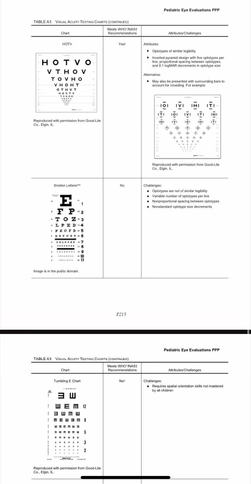
0-5岁检查眼部项目推荐表

-
眼结膜下出血 Conjunctival hemorrhage
眼结膜下出血系由于结膜小血管破裂。 见于外伤和眼部手术后,更为常见的是自发性出血。 见于严重 急性结膜炎 的结膜下出血,可能是由微小 血管栓塞 所致。 自发出血多见于老年人、背驮重物、恶心呕吐、剧烈咳嗽、喷嚏、便秘等诱因,亦可致小 …
别称: 眼结膜下出血
中医病名: 眼结膜下出血
英文名称: Conjunctival hemorrhage
就诊科室: 眼科
· 发病原因· 眼结膜下出血系由于结膜小血管破裂。见于外伤和眼部手术后,更为常见的是自发性出血。见于严重急性结膜炎的结膜下出血,可能是由微小血管栓塞所致。自发出血多见于老年人、背驮重物、恶心呕吐、剧烈咳嗽、喷嚏、便秘等诱因,亦可致小血管: 破裂出血。坏血症、各种血液病、紫癜、动脉硬化、糖尿病、高血压以及疟疾等高热性传染病亦可发生。
· 当颅骨底部骨折后,出血沿眶底向前扩延到下穹隆部、下方球结膜及下睑皮下,出血量一般较大。蝶骨骨折晚期出血出现在颞侧较其他部位多。一般这种出血多发生在头颅外伤后,是颅底骨折重要体征之一。
· 严重眼球钝挫伤,结膜下出血量大,呈黑红色者应注意排除巩膜破裂之可能。结膜下出血呈鲜纡色。平坦、境界清楚、点或片状,出血量多时呈黑红色。除急性结膜炎引起的出血外,不伴炎症体征。出血逐渐吸收,颜色由红色变为棕、黄色雨消失,不留痕迹。局部无需治疗,通常1~3周可吸收消退。反复出血者应查找致病原因,予以治疗。
· 发病
· 1、高血压∶好发于老年人,若合并“结膜下出血”,应定期测量血压,其他如糖尿病或心血管疾病患者也易引发,需加以留意。
· 2、急性结膜炎∶由70型肠病毒所引起,具高度传染性,若合并结膜水肿,眼皮肿胀,分泌物增加时需高度怀疑。
· 3、局部轻微受伤引起∶例如因过敏性结膜炎或干眼症用力揉擦眼睛所造成;需闭气用力的便秘解便、生产、搬重物等;以及伤风感冒、被辛辣食物呛到而导致咳嗽的情况。
胺碘酮,普罗帕酮致眼结膜下出血
何满仓,刘莉摘要:胺碘酮(可达龙),自1961年问世以来,开始用于治疗心绞痛,现已广泛用于治疗各种快速型心律失常,普罗帕酮为Ic类抗心律失常药,对冠心病、高血压所引起的心律失常有较好的疗效.现报告2药致眼结膜下出血1例如下.
关键词:普罗帕酮 胺碘酮 眼结膜下出血 抗心律失常药 小陇山林业实验局 冠心病 交界性早搏 药物不良反应 高血压 心绞痛
Subconjunctival Hemorrhage
Medical Condition
Breakage of small blood vessel below the clear surface of the eye.
Common (More than 200,000 cases per year in US)
Doesn't require lab test or imaging
Can last several days or weeks
Mainly due to breakage of small, delicate blood vessels behind the conjunctiva due to sudden pressure or straining. Characterized by a bright red patch on the sclera of the eye which is painless but have a scratchy feeling. In most cases, no treatment is required for this condition, mainly supportive care and monitoring for changes and improvement.
Treatments
In most cases, no treatment is required for this condition, mainly supportive care and monitoring for changes and improvement.Medication
Artificial tears: To relieve irritation.
Ocular lubricant
Antibiotics: Topical antibiotics in presence of infection.
Tetracycline · Neomycin
Self careAvoid redness reliever eye drops
Avoid rubbing the eye
Proper nutrition
See the doctor immediately if it appears in both the eyes
People also search for
Red EyeHyphema
Breakage of small blood vessel below the clear surface of the eye.
Common (More than 200,000 cases per year in US)
发病率每年在美国20万例结膜出血
Doesn't require lab test or imaging
不需要特殊实验室检查或影像检查
Can last several days or weeks
持续数日数周,实际我碰到的激光手术后结膜出血持续3-5周。
Common for ages 50 and older
年龄通畅50岁或以上
More common in males男性更常见结膜出血
Subconjunctival hemorrhage (broken blood vessel in eye ...
https://www.mayoclinic.org/diseases-conditions/sub...A subconjunctival hemorrhage (sub-kun-JUNK-tih-vul HEM-uh-ruj) occurs when a tiny blood vessel breaks just underneath the clear surface of your eye (conjunctiva). In many ways, it's just like having a bruise on your skin. The conjunctiva can't absorb blood very quickly, so the blood gets trapped. You may not even realize you have a subconjunctival hemorrhage until you look in the mirror and notice that the white paart of your eye is bright red.
A subconjunctival hemorrhage often occurs without any obvious harm to your eye. Even a strong sneeze or cough can cause a blood vessel to break in the eye. You don't need to treat it. A subconjunctival hemorrhage may look alarming, but it's usually a harmless condition that disappears within two weeks or so.
Broken blood vessel in the eyeA subconjunctival hemorrhage often occurs without any obvious harm to your eye. Even a strong sneeze or cough can cause a blood vessel to break in the eye. You don't need to treat it. A subconjunctival hemorrhage may look alarming, but it's usually a harmless condition that disappears within two weeks or so.
Products & Services
· Book: Mayo Clinic Guide to Better VisionSymptoms
The most obvious sign of a subconjunctival hemorrhage is a bright red patch on the white (sclera) of your eye.Despite its bloody appearance, a subconjunctival hemorrhage looks worse than it is and should cause no change in your vision, discharge or pain. Your only discomfort may be a scratchy feeling on the surface of the eye.
When to see a doctor
If you have recurrent subconjunctival hemorrhages or other bleeding, talk to your doctor.结膜下再次或反复出血或其他部位同时出血需要咨询专业的医生Causes
The cause of a subconjunctival hemorrhage isn't always known. The following actions may cause a small blood vessel to rupture in your eye:· Violent coughing
· Powerful sneezing
· Straining
· Vomiting
In some cases, a subconjunctival hemorrhage may result from an eye injury, including:
· Roughly rubbing your eye
· Trauma, such as a foreign object injuring your eye
Risk factors
Risk factors for a subconjunctival hemorrhage include:· Diabetes
· High blood pressure (hypertension)
· Certain blood-thinning medications, such as warfarin (Coumadin, Jantoven) and aspirin
· Blood-clotting disorders
Complications
Health complications from a subconjunctival hemorrhage are rare. If your condition is due to trauma, your doctor may evaluate your eye to ensure you don't have other eye complications or injury.Prevention
If the bleeding on the surface of your eye has a clearly identifiable cause, such as a bleeding disorder or blood-thinning medication, ask your doctor if you can take any steps to reduce the risk of a subconjunctival hemorrhage.If you need to rub your eyes, rub them gently. Rubbing too hard can cause minor trauma to your eyes, which may lead to a subconjunctival hemorrhage.
Sept. 21, 2021
Related
Associated Procedures
Blood pressure test
Eye exam
Products & Services
Book: Mayo Clinic Guide to Better Vision
Subconjunctival hemorrhage (broken blood vessel in eye)
Symptoms & causes
Diagnosis & treatment
Doctors & departments
-

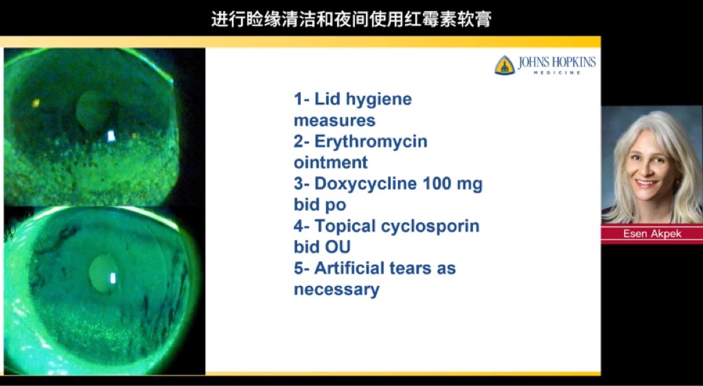
Hordeolum是一种在眼睑中发现的急性细菌感染。[1]这种感染是一种常见疾病,患者经常到他们的初级保健医生或急性护理中心进行评估和治疗。[2]患者通常表现为眼睑疼痛性红斑性炎症。麦粒体可以在外眼睑上形成,通常被称为麦粒肿。它也可能在内眼睑上形成,很容易被误认为是霰粒肿。[3]这种情况通常持续一到两周,是自限性的,并且经常自行消退。可以用热敷和按摩疗法治疗。可能需要局部使用抗生素,在极少数情况下,脓疱可能需要引流。[3]
病因学
它通常是由感染睫毛毛囊的葡萄球菌引起的。外部麦粒肿是由皮脂腺(蔡司)腺或汗腺(毛细胞)腺的阻塞引起的。[4]阻塞发生在睫毛线处,表现为疼痛的红色肿胀区域,发展成脓疱。内部麦粒肿是由睑板腺阻塞引起的,脓疱在眼睑的内表面形成。[3] Hordeola可能同时出现在上眼睑和下眼睑上。[4]
流行病学
Hordeolum是家庭实践和急性护理环境中的常见表现。[2]种族,性别或性别与大麦病流行率之间没有直接相关性。由于皮脂粘度增加,成人可能更容易患病。患有睑缘炎,脂溢性皮炎,酒渣鼻,糖尿病和脂质升高等疾病的患者也面临发生群淋巴细胞的风险增加。[3]
病理 生理
感染是由于蔡司、摩尔或睑板腺分泌物增厚、干燥或淤滞引起的。蔡司和莫尔腺是眼睛的睫状体。蔡司腺分泌具有防腐特性的皮脂,可以防止细菌的生长。[2]摩尔腺产生免疫球蛋白A,粘蛋白1和溶酶体,这对于免疫防御眼睛中的细菌至关重要。[5]当这些腺体堵塞或阻塞时,眼睛防御就会受损。淤滞可导致细菌感染,金黄色葡萄球菌是最常见的病原体。[4]在白细胞浸润发生局部炎症反应后,会出现化脓性口袋或脓肿。
历史和物理
仔细的病史和体格检查至关重要。患者通常会缓慢而隐匿地出现疼痛、发红和肿胀的眼睑,无异物或外伤史。如果麦粒肿的大小压在角膜上,视力可能会受到影响。患者不应报告眼痛,眼外运动应完整无痛。红斑局限于患眼睑。提供者应尝试定位脓疱,并且可能需要切除眼睑部分局限影响引流形成纤维包膜,特别是定位内部的麦粒肿。提供者应询问麦粒肿的任何诱发性疾病,这些疾病应在治疗中得到解决。眼球运动伴眶周肿胀和红斑的任何疼痛均提示眼眶蜂窝织炎,需要额外和更积极的管理和治疗。眼部持续或复发性疼痛肿块可能提示癌,需要活检。在这些情况下需要转诊眼科。[2][6][3]
评估
通常,没有与麦粒肿相关的诊断性检查,这是一种临床诊断。在极少数情况下,如果发生并发症,并且感染扩散并导致眶周或眼眶蜂窝织炎,则需要额外的检查和影像学检查。有时,内部麦粒肿会引起角膜刺激,在这种情况下,提供者可能会用荧光素染色眼睛,以确保没有角膜擦伤。[2]
治疗/管理
在许多情况下,病变可以在没有任何治疗的情况下自发引流。热敷也是有益的,按摩该区域也是如此。热敷的目的是软化肉芽肿组织并促进引流。迄今为止,还没有结论性研究表明,仅这种方法就会导致持续时间缩短或结局改善。眼睑按摩旨在帮助表达受感染腺体的脓性引流。使用无泪且 PH 平衡的生理盐水或温和不刺激角膜的洗面奶(例如婴儿洗面奶)进行眼睑清洁,可通过清除堵塞导管中的碎屑来促进排出。肥皂还可通过分解细胞膜来帮助去除细菌,它还可以治疗外部麦粒肿的根本原因,睑缘炎。[1]应特别注意挤压和按摩内部麦粒肿,因为这可能会导致角膜刺激或变形。[7]持续性病变或较大病变可能需要抗生素治疗。这种治疗可能有助于缩短持续时间和严重程度。经常使用大环内酯类抗生素眼膏,如红霉素眼药膏,并具有润滑的额外好处。如果肿胀明显并对角膜造成压力,则可以短期使用局部类固醇。[8]如果感染扩散并进展为眶周或眼眶蜂窝织炎,则需要全身性抗生素。[2]持续性脓肿的切口和引流可能是必要的。[9]眼科医生应在局部麻醉下进行切口和引流。应将标本送往病理学,以排除更严重的疾病,包括癌症。
鉴别诊断
基底细胞癌
霰粒肿
气肿-眼眶(罕见)
前期蜂窝织炎
皮脂腺癌
鳞状细胞癌
形成颗粒和其他问题
虽然麦粒肿是一种常见的表现,但医生应确保在评估和治疗期间考虑并排除红眼皮疼痛的其他表现。其他应考虑的诊断包括眶周和眼眶蜂窝织炎、霰粒肿、皮脂腺癌和鳞状细胞癌。[6]提供者还应考虑可能导致群病复发的根本原因,如睑缘炎和酒渣鼻。应解决这些基础疾病,以防止这些患者群体中麦粒肿复发。[10]霰粒肿可以模仿内部的Hordeolum,起初可能很难区分两者。霰粒肿在眼睑中间的皮脂腺周围形成。它是由腺体中泄漏到周围组织的分泌物分解形成的。最初,炎症可能产生疼痛,并可能表现为内部麦粒肿。然而,霰粒肿发展为无痛肉芽肿性结节,被认为是无菌性慢性炎症。[8]
提高医疗团队的成果
Hordeolum经常由急诊医生,执业护士或初级保健提供者遇到。在大多数情况下,感染可以通过保守治疗来控制。热敷的目的是软化肉芽肿组织并促进引流。持续性病变或较大病变可能需要抗生素治疗。这种治疗可能有助于缩短持续时间和严重程度。但是,如果感染量较大或病因不明,应将患者转诊至眼科医生处。[9]眼科医生应在局部麻醉下进行切口和引流。应将标本送往病理学,以排除更严重的疾病,包括癌症。对于大多数大麦病患者,结果非常好。
A hordeolum is an acute, common bacterial infection of the eyelid. Patients with this condition often present to their primary care provider with painful, erythematous inflammation of the eyelid. This activity illustrates the evaluation and treatment of hordeolum and reviews the role of the professional team in managing patients with this condition.
Objectives:
Describe the typical clinical presentation of a hordeolum.
Review the pathophysiology of hordeolum.
Summarize the use of warm compresses, lid massage, and lid scrubs in the management of hordeolum.
Identify the importance of improving care coordination among the interprofessional team to improve outcomes for patients affected with hordeolum.
Access free multiple choice questions on this topic.
Introduction
A hordeolum is an acute bacterial infection found in the lid of the eye.[1] This infection is a common condition, and patients often present to their primary care physician or acute care center for evaluation and treatment.[2] The patient usually presents with painful, erythematous inflammation of the eyelid. The hordeolum can form on the external eyelid and is referred to commonly as a stye. It may also form on the inner eyelid and can be easily mistaken for a chalazion.[3] The condition often lasts one to two weeks, is self-limiting, and often resolves on its own. It may be treated with warm compresses and massage therapy. Topical antibiotics may be indicated, and in rare cases, the pustule may require drainage.[3]
Etiology
It is usually caused by Staphylococcus that infects the eyelash hair follicle. The external hordeolum is caused by a blockage of the sebaceous (Zeis) glands or sweat (Moll) glands.[4] The blockage occurs at the lash line and presents as a painful red swollen area that develops into a pustule. The internal hordeolum is caused by a blockage of the Meibomian glands, and the pustule forms on the inner surface of the eyelids.[3] Hordeola may present on both the upper and the lower eyelids.[4]
Epidemiology
Hordeolum is a common presentation in family practice and acute care settings.[2] There is no direct correlation between race, sex, or gender with regards to hordeolum prevalence. Adults may be more prone due to the increased viscosity of the sebum. Patients with conditions such as blepharitis, seborrheic dermatitis, rosacea, diabetes, and elevated lipids are also at increased risk for the development of hordeola.[3]
Pathophysiology
The infection occurs due to thickening, drying, or stasis of the Zeis, Moll, or Meibomian gland secretions. The Zeis and Moll glands are the ciliary glands of the eye. The Zeis gland secretes sebum with antiseptic properties that may prevent the growth of bacteria.[2] The Moll gland produces immunoglobulin A, mucin 1, and lysosomes which are essential in the immune defense against bacteria in the eye.[5] When these glands become clogged or blocked, the eye defenses are impaired. The stasis can lead to bacterial infection with Staphylococcus aureus being the most common pathogen.[4] After a localized inflammatory response occurs with infiltration by leukocytes, a purulent pocket or abscess develops.
History and Physical
A careful history of and physical exam is essential. The patient will usually relay a slow and insidious onset of a painful, red, and swollen eyelid without a history of foreign body or trauma. Visual acuity may be affected if the size of the hordeolum is pressing on the cornea. The patient should not report ocular pain, and their extraocular movements should be intact and painless. The erythema is localized to the lid of the affected eye. The provider should try to locate a pustule, and the eyelids may need to be everted, especially to locate an internal hordeolum. The provider should inquire about any of the predisposing conditions for hordeolum, and these conditions should be addressed and managed in treatment. Any pain in ocular movements with periorbital swelling and erythema is indicative of orbital cellulitis and requires additional and more aggressive management and treatment. Persistent or recurrent painful lumps in the eye may be indicative of carcinoma and require biopsy. Ophthalmology referral is indicated in these situations.[2][6][3]
Evaluation
Typically, there is no diagnostic testing associated with a hordeolum, and it is a clinical diagnosis. Rarely, additional testing and imaging will be required if complications occur, and the infection spreads and causes periorbital or orbital cellulitis. Occasionally, an internal hordeolum can cause corneal irritation, in which case the provider may stain the eye with fluorescein to ensure there is no corneal abrasion.[2]
Treatment / Management
In many cases, the lesions can spontaneously drain without any treatment. Warm compresses are also of benefit, as is massage to the area. These are often seen as the gold standard. Warm compresses are aimed at softening the granulomatous tissue and facilitating drainage. There are no conclusive studies to date, showing that this method alone causes any shortened durations or improved outcomes. Lid massage is intended to help express the purulent drainage from the infected gland. Lid scrubs with saline or mild shampoo (e.g., baby shampoo) that is tear-free and ph-balanced, may promote drainage by clearing debris from the clogged duct. Soap may also help to remove bacteria by breaking down cell membranes, and it may also treat an underlying cause of the external hordeolum, blepharitis.[1] Careful attention should be paid to compresses and massage for the internal hordeolum, as this could cause irritation or deformation to the cornea.[7]Persistent lesions or larger lesions may require antibiotic therapy. This treatment may help to shorten duration and severity. A macrolide antibiotic ointment such as erythromycin ophthalmic ointment is often used and has the added benefit of lubrication. If the swelling is significant and causing pressure on the cornea, topical steroids can be used for a short duration.[8] If the infection spreads and progresses to a periorbital or orbital cellulitis, systemic antibiotics are required.[2] Incision and drainage of a persistent abscess may be necessary.[9] An ophthalmologist should perform the incision and drainage under local anesthesia. The specimen should be sent to pathology to rule out more serious diseases, including carcinoma.
Differential Diagnosis
Basal cell carcinoma
Chalazion
Pneumo-Orbita(rare)
Preseptal cellulitis
Sebaceous gland carcinoma
Squamous cell carcinoma
Pearls and Other Issues
While hordeolum is a common presentation, the practitioner should ensure that other manifestations of a painful red eyelid are considered and ruled out during evaluation and treatment. Other diagnoses that should be considered are periorbital and orbital cellulitis, chalazion, sebaceous gland carcinoma, and squamous cell carcinoma.[6] The provider should also consider underlying causes that can lead to a reoccurrence of hordeola such as blepharitis and rosacea. These underlying conditions should be addressed to prevent recurrent hordeolum in these patient populations.[10]A chalazion can mimic an internal hordeolum, and it may be difficult to distinguish between the two at first. The chalazion forms around the sebaceous gland in the middle of the eyelid. It forms from the breakdown of the secretions in the gland that leak into the surrounding tissues. Initially, the inflammation may produce pain and may present as an internal hordeolum. However, the chalazion develops into a painless granulomatous nodule and is considered an aseptic, chronic inflammation.[8]
Enhancing Healthcare Team Outcomes
Hordeolum is often encountered by the emergency physician, nurse practitioner, or primary care provider. In most cases, the infection can be managed with conservative treatment. Warm compresses are aimed at softening the granulomatous tissue and facilitating drainage. Persistent lesions or larger lesions may require antibiotic therapy. This treatment may help to shorten duration and severity. However, if the infection is large or the cause is not known, the patient should be referred to an ophthalmologist.[9] An ophthalmologist should perform the incision and drainage under local anesthesia. The specimen should be sent to pathology to rule out more serious diseases, including carcinoma.The outcomes for most patients with hordeolum is excellent.
Review Questions
References
1.Lindsley K, Nichols JJ, Dickersin K. Non-surgical interventions for acute internal hordeolum. Cochrane Database Syst Rev. 2017 Jan 09;1:CD007742. [PMC free article] [PubMed]
2.Pflipsen M, Massaquoi M, Wolf S. Evaluation of the Painful Eye. Am Fam Physician. 2016 Jun 15;93(12):991-8. [PubMed]
3.McAlinden C, González-Andrades M, Skiadaresi E. Hordeolum: Acute abscess within an eyelid sebaceous gland. Cleve Clin J Med. 2016 May;83(5):332-4. [PubMed]
4.Wald ER. Periorbital and orbital infections. Pediatr Rev. 2004 Sep;25(9):312-20. [PubMed]
5.Takahashi Y, Watanabe A, Matsuda H, Nakamura Y, Nakano T, Asamoto K, Ikeda H, Kakizaki H. Anatomy of secretory glands in the eyelid and conjunctiva: a photographic review. Ophthalmic Plast Reconstr Surg. 2013 May-Jun;29(3):215-9. [PubMed]
6.Carlisle RT, Digiovanni J. Differential Diagnosis of the Swollen Red Eyelid. Am Fam Physician. 2015 Jul 15;92(2):106-12. [PubMed]
7.McMonnies CW, Korb DR, Blackie CA. The role of heat in rubbing and massage-related corneal deformation. Cont Lens Anterior Eye. 2012 Aug;35(4):148-54. [PubMed]
8.Jin KW, Shin YJ, Hyon JY. Effects of chalazia on corneal astigmatism : Large-sized chalazia in middle upper eyelids compress the cornea and induce the corneal astigmatism. BMC Ophthalmol. 2017 Mar 31;17(1):36. [PMC free article] [PubMed]
9.Hirunwiwatkul P, Wachirasereechai K. Effectiveness of combined antibiotic ophthalmic solution in the treatment of hordeolum after incision and curettage: a randomized, placebo-controlled trial: a pilot study. J Med Assoc Thai. 2005 May;88(5):647-50. [PubMed]
10.Papier A, Tuttle DJ, Mahar TJ. Differential diagnosis of the swollen red eyelid. Am Fam Physician. 2007 Dec 15;76(12):1815-24. [PubMed] -

LipiFlow
Johnson and Johnson Vision make the LipiFlow thermal pulsation system. It applies heat and pressure to melt the waxy and oily deposits clogging meibomian glands, allowing the glands to express their oils healthily and improving the quality and quantity of the lipid layer.
This 12-minute in-office treatment can improve MGD and dry eye symptoms for up to three years.
iLUX
The in-office iLUX treatment uses an LED heat source to melt the waxy oil clogging your glands. Then the eye doctor will apply pressure to help express the clogged glands.
This treatment is manufactured by Tear Film Innovations and won Gold at the 2019 Medical Design Excellence Awards (MDEA ).
TearCare
Sight Sciences developed TearCare. It uses adhesive heating patches connected to a small heating unit to warm the waxy deposits. Then forceps press your lids and glands open to begin proper expression.
Intense Pulsed Light (IPL )
IPL treatment has been used by dermatologists for years to treat acne rosacea.
It has also shown to be successful in alleviating MGD and dry eye symptoms. IPL shines intense flashes of infrared and visible light on the eyelids over 20 minutes. This is done in multiple treatment sessions about a month apart.
Blephex
Blephex treats blepharitis and MGD through exfoliation.
A hand-held instrument with a small rotating sponge attached applies pressure and scrubs the conjunctiva (eyelid margin ). The sponge gently exfoliates and removes inflammatory biofilm that clogs your glands. The process takes about 10 minutes.
Lid Debridement
Mechanical lid debridement involves using tools to scrape the eyelid to remove keratin and other debris from the meibomian gland orifices around the lid margins.
Antibacterials
Some eye doctors may prescribe you antibacterial eye drops in order to restore proper meibomian gland function. This includes topical antibiotics such as the gel azithromycin.
They may also prescribe systemic antibiotics such as doxycycline or ertythromycin.
Cyclosporine Eye Drops
Cyclosporine (brand names: Restasis and Cequa ) modifies the body’s immune response. It increases your ability to produce more tears, making it a treatment option for dry eyes and MGD.
Prescription Medications
There are no medications that are approved by the FDA to treat MGD, however, there are two medications Xiidra is FDA approved for the treatment of dry eye. It may be used off-label to alleviate symptoms of MGD.
Omega-3 Supplements
Some eye doctors suggest that omega-3 fatty acids may provide a useful adjunct treatment for MGD. These essential fatty acids may help suppress conjunctival inflammation and decrease the risk of future problems.
-

眼睛干涩
眼睛干燥等,当眼泪无法稳定保持眼表充分润滑时,就会发生这种情况。常见原因:
老花
药物
激光眼科手术
干燥空气过多
减少眨眼
长时间使用计算机
隐形眼镜的使用
女性激素变化
治疗
自我治疗:在一些不太严重的情况下可能有帮助:
使用防护眼镜阻挡干燥空气
加湿室内空气
将计算机屏幕置于视线水平以下
避免接触烟雾
使用人工泪液
通过眨眼和闭上眼睛来频繁地锻炼眼睛
富含维生素A食物和不饱和脂肪酸适当补充(肝脏,胡萝卜和西兰花)欧米伽脂肪酸-3, EPA, DHA
如果您发现以下情况,请去看医生:
眼睛发红
眼睛疼痛
阅读困难
如果您发现以下情况,请立即就医:
视力丧失
视力
感染的迹象,如发烧,炎症,液体排出可用于确定干眼症原因的测试和程序包括:1.全面的眼科检查。眼科检查包括您的整体健康状况和眼睛健康的完整病史,可以帮助您的医生诊断您眼睛干涩的原因;特别是角膜荧光素染色下的泪液破裂时间检查(BUT检查,角膜荧光素是否染色阳性);2.测量眼泪量的测试。您的医生可能会使用Schirmer试验来测量您的泪液产生。结膜印迹细胞试验...3.泪液渗透压检查 4.睑板腺相关检查:分布,开头透明度,堵塞等检查和评估等
Diagnosis
Tests and procedures that may be used to determine the cause of your dry eyes include:A comprehensive eye exam. An eye exam that includes a complete history of your overall health and your eye health can help your doctor diagnose the cause of your dry eyes.
A test to measure the volume of your tears. Your doctor may measure your tear production using the Schirmer test. In this test, blotting strips of paper are placed under your lower eyelids. After five minutes your doctor measures the amount of strip soaked by your tears.
Another option for measuring tear volume is the phenol red thread test. In this test, a thread filled with pH-sensitive dye (tears change the dye color) is placed over the lower eyelid, wetted with tears for 15 seconds and then measured for tear volume.
A test to determine the quality of your tears. Other tests use special dyes in eyedrops to determine the surface condition of your eyes. Your doctor looks for staining patterns on the corneas and measures how long it takes before your tears evaporate.
A tear osmolarity test. This type of test measures the composition of particles and water in your tears. With dry eye disease, there will be less water in your eyes.
Tear samples to look for markers of dry eye disease, including elevated matrix metallo proteinase-9 or decreased lactoferrin.
More Information
Eye exam
Treatment
Punctal plugsOpen pop-up dialog box
For most people with occasional or mild dry eye symptoms, it's enough to regularly use over-the-counter eyedrops (artificial tears). If your symptoms are persistent and more serious, you have other options. What you do depends on what's causing your dry eyes.Some treatments focus on reversing or managing a condition or factor that's causing your dry eyes. Other treatments can improve your tear quality or stop your tears from quickly draining away from your eyes.
Treating the underlying cause of dry eyes
In some cases, treating an underlying health issue can help clear up the signs and symptoms of dry eyes. For instance, if a medication is causing your dry eyes, your doctor may recommend a different medication that doesn't cause that side effect.If you have an eyelid condition, such as your lids turned outwards (ectropion), your doctor may refer you to an eye surgeon who specializes in plastic surgery of the eyelids (oculoplastic surgeon).
Medications
Prescription medications used to treat dry eyes include:Drugs to reduce eyelid inflammation. Inflammation along the edge of your eyelids can keep oil glands from secreting oil into your tears. Your doctor may recommend antibiotics to reduce inflammation. Antibiotics for dry eyes are usually taken by mouth, though some are used as eyedrops or ointments.
Eyedrops to control cornea inflammation. Inflammation on the surface of your eyes (cornea) may be controlled with prescription eyedrops that contain the immune-suppressing medication cyclosporine (Restasis) or corticosteroids. Corticosteroids are not ideal for long-term use due to possible side effects.
Eye inserts that work like artificial tears. If you have moderate to severe dry eye symptoms and artificial tears don't help, another option may be a tiny eye insert that looks like a clear grain of rice. Once a day, you place the hydroxypropyl cellulose (Lacrisert) insert between your lower eyelid and your eyeball. The insert dissolves slowly, releasing a substance that's used in eyedrops to lubricate your eye.
Tear-stimulating drugs. Drugs called cholinergics (pilocarpine, cevimeline) help increase tear production. These drugs are available as pills, gel or eyedrops. Possible side effects include sweating.
Eyedrops made from your own blood. These are called autologous blood serum drops. They may be an option if you have severe dry eye symptoms that don't respond to any other treatment. To make these eyedrops, a sample of your blood is processed to remove the red blood cells and then mixed with a salt solution.
Other procedures
Other procedures that may be used to treat dry eyes include:Closing your tear ducts to reduce tear loss. Your doctor may suggest this treatment to keep your tears from leaving your eye too quickly. This can be done by partially or completely closing your tear ducts, which normally serve to drain tears away.
Tear ducts can be plugged with tiny silicone plugs (punctal plugs). These are removable. Or tear ducts can be plugged with a procedure that uses heat. This is a more permanent solution called thermal cautery.泪点塞
Using special contact lenses. Ask your doctor about newer contact lenses designed to help people with dry eyes.Some people with severe dry eyes may opt for special contact lenses that protect the surface of the eyes and trap moisture. These are called scleral lenses or bandage lenses.巩膜镜或绑带镜
Unblocking oil glands. Warm compresses or eye masks used daily can help clear up blocked oil glands. A thermal pulsation device is another way to unclog the oil glands, but it is unclear whether this method provides any advantage over warm compresses.
Using light therapy and eyelid massage. A technique called intense-pulsed light therapy followed by massage of the eyelids has proved to help people with severe dry eyes.
Lifestyle and home remedies
You may be able to manage your dry eyes with frequent eyelid washing and use of over-the-counter (OTC) eyedrops or other products that help lubricate your eyes. If your condition is long term (chronic), use eyedrops even when your eyes feel fine to keep them well lubricated.Selecting and using OTC products for dry eyes
A variety of nonprescription products for dry eyes are available, including eyedrops (artificial tears), gels and ointments. Talk with your doctor about which might be best for you.Artificial tears may be all you need to control mild dry eye symptoms. Some people need to put drops in several times a day, and some use them only once a day.
Consider these factors when selecting an OTC product:
Preservative vs. nonpreservative drops. Preservatives are added to some eyedrops to prolong shelf life. You can use eyedrops with preservatives up to four times a day. But using the preservative drops more often can cause eye irritation.
Nonpreservative eyedrops come in packages that contain multiple single-use vials. After you use a vial, you throw it away. If you rely on eyedrops more than four times a day, nonpreservative drops are safe.
Drops vs. ointments. Lubricating eye ointments coat your eyes, providing longer lasting relief from dry eyes. But these products are thicker than eyedrops and can cloud your vision. For this reason, ointments are best used just before bedtime. Eyedrops can be used at any time and won't interfere with your vision.
Drops that reduce redness. It's best to avoid these as your solution for dry eyes, as prolonged use can cause irritation.
Washing your eyelids to control inflammation
For people with blepharitis and other conditions that cause eyelid inflammation that blocks the flow of oil to the eye, frequent and gentle eyelid washing may help. To wash your eyelids:Apply a warm washcloth to your eyes热敷. Wet a clean cloth with warm water. Hold the cloth over your eyes for five minutes. Rewet the cloth with warm water when it cools. Gently rub the washcloth over your eyelids — including the base of the eyelashes — to loosen any debris.
Use a mild soap on your eyelids温和的肥皂. Use baby shampoo or another mild soap. Put the cleanser on your clean fingertips and gently massage your closed eyes near the base of your eyelashes. Rinse completely.
More Information
Artificial tears人工泪液选择: How to select eyedrops for dry eyes
Alternative medicine替代治疗
Further study is needed, but some alternative medicine approaches may help relieve your dry eye symptoms. Discuss the benefits and risks with your doctor.Fatty acids. omega-3脂肪酸口服Adding omega-3 fatty acids to your diet may help relieve dry eye signs and symptoms. These are available as supplements and in foods such as flaxseed, salmon and sardines.
Castor oil eyedrops蓖麻油滴眼液. These eyedrops may improve symptoms by reducing tear evaporation.
Acupuncture针灸. Some people have seen their dry eye symptoms improve after acupuncture therapy.
Preparing for your appointment
You're likely to start by seeing your family doctor. He or she may then refer you to an eye specialist (ophthalmologist). Because appointments can be brief, it's a good idea to be well prepared for your appointment.What you can do
List any symptoms you're experiencing, including any that may seem unrelated to the reason for which you scheduled the appointment.
List key personal information, including any recent life changes.
Make a list of all medications, vitamins and supplements that you're taking.
List questions to ask your doctor.
For dry eyes, some basic questions to ask your doctor include:What's the most likely cause of my dry eyes?
Do I need any tests?
Can dry eyes get better on their own?
What are my treatment options?
What are the potential side effects of each treatment?
I have other health conditions. How can I best manage these conditions together?
Is a generic drug available for the medicine you're prescribing me?
Do you have any brochures or other printed material that I can take with me?
What websites do you recommend?
Do I need to plan for a follow-up visit?
Don't hesitate to ask additional questions that may occur to you during your appointment.What to expect from your doctor
Your doctor may ask:Can you describe your symptoms?
Do you recall when you first began experiencing symptoms?
Have your symptoms been continuous or occasional?
Do other members of your family have dry eyes?
Have you tried over-the-counter eyedrops? Did they provide relief?
Are your symptoms worse in the morning or late in the day?
What medications do you take?
Have you had any radiation to the head or neck?
What you can do in the meantime
To relieve your signs and symptoms while you wait for your appointment, try over-the-counter eyedrops. Look for lubricating eyedrops (artificial tears) and avoid those that advocate reducing redness in the eyes. Eyedrops that reduce eye redness can cause additional eye irritation.
Sept. 24, 2020
Book: Mayo Clinic Guide to Better Vision -
 Contents 内容• 1 Introduction 介绍• 2 Evaluation 评估• 3 Treatment 治疗• 4 Post Treatment 随访• 5 References 参考文献Introduction角膜异物(FB)是一种表面附着或嵌在角膜内的物体。角膜是眼球的最前部,接触异物最多。一些常见的可能嵌入角膜的异物包括玻璃、金属、沙子、塑料或木材。摘除角膜异物是在办公室或急诊室进行的。症状包括异物感、疼痛、流泪、光敏感和视力下降。[1][2]A corneal foreign body (FB) is an object that is superficially adherent or embedded in the cornea. As the most anterior part of the globe, the cornea is the most exposed to foreign bodies. Some of the common foreign bodies that may be embedded in the cornea include glass, metal, sand, plastic, or wood. The removal of a corneal foreign body is typical performed in an office or emergency room setting. Symptoms include foreign body sensation, pain, tearing, light sensitivity and decreased vision.[1][2]EvaluationShould any foreign body become lodged in the cornea, it is important to obtain a thorough history to prepare for the procedure and provide appropriate patient care. Some questions to ask include: What? When? Where and How? For example, an iron foreign body will start forming a rust ring after four to six hours of being lodged in the cornea. Injuries caused by vegetable matter or soil are more likely to get infected. Knowing the mechanism of the injury is important in eliciting the force with which the FB entered the corneal and determining the need for any additional tests to evaluate for possible ocular perforation and intraocular foreign bodies.[3] Some of those tests include an ocular ultrasound (B scan), thin cut orbital CT scan, and gonioscopy. Patient might need an updated tetanus immunization.[4]After obtaining detailed history, patient should have their best corrected visual acuity checked, followed by a thorough slit lamp exam. Assess the type and depth of the FB carefully. Deeply embedded FBs may need to be removed in the OR. Fluorescein dye test is beneficial to highlight a FB and any corneal abrasions. [3] Fornices should be everted to look for any additional FBs.[2]Extreme caution must be exercised in order to remove corneal FBs safely and in timely fashion to minimize risk of infection, inflammation, scarring, and vision loss. [3] With time, the foreign body can get pushed deeper and deeper into the cornea, sometimes penetrating all the way through the entire cornea and becoming dislodged internally within the eye. Glass and fiberglass FBs are generally well tolerated in the corneal stroma and can occasionally be monitored if the removal will cause more damage. [5]Treatment患者的眼表应采用局部麻醉,如0.5%盐酸丙帕卡因或0.5%盐酸丁卡因,使治疗对患者没有任何额外的疼痛或不适。给病人一个目标以保持眼睛稳定。上眼睑和下眼睑可以用指尖轻轻固定,基本上固定眼睑。在大多数情况下,针是最好的工具。一个25或27号的5/8“针是一个很好的选择,因为它提供了足够的力量将FB从角膜表面提起。[2][3]通过裂隙灯观察,以斜向或切向角入近角膜,以避免角膜穿孔。拧紧FB的边缘并松开它。人们可以使用一个微妙的摆动动作来完成这个过程。异物松动时,可以用镊子轻轻从眼睛中取出。[3]在涉及植物物质、长针状异物或异物附着在角膜表面的病例中,珠宝商钳可能是摘除的首选工具。金属异物可以用磁性开钻去除。[3]一种无菌湿棉尖可用于去除表面异物。偶尔,结膜囊冲洗被用来清除多个小颗粒。[2][4]在去除金属异物后,可能会留下一个棕橙色的铁锈环。它可以用针或镊子举起。大多数情况下使用电刷去除除锈环。其目标是尽可能多地清除锈,而不会造成太多的组织破坏或角膜穿孔。移除角膜异物后,重新评估角膜是否有残留的外来颗粒,评估挖掘的深度和上皮缺损的范围。用荧光素钠染料和钴蓝光进行最终评价。[3]最终塞德尔测试表明,在深残留缺陷的情况下。治疗后摘除异物后,患者应使用广谱局部眼科抗生素一周或直到角膜表面再上皮化。一种治疗性绷带,隐形眼镜可以短期使用,以减少不适。晶状体作为一种屏障,减少眼睑对角膜表面的剪切力,最大限度地减少上皮细胞撕裂的风险,并促进愈合。绷带隐形眼镜应谨慎使用,因为它可以促进更传染性的环境,应密切监测。短效局部睫状体麻痹滴剂可用于减轻不适。[2]在切除角膜异物后,患者通常应在24小时内就诊,以评估角膜是否有任何发展中的感染、水肿和上皮缺损。确切的随访和术后护理将取决于角膜异物的性质和深度。[3]The patient’s ocular surface should be anesthetized with topical anesthetic such as ophthalmic proparacaine hydrochloride 0.5% or tetracaine hydrochloride 0.5% to allow the treatment to be performed without any additional pain or discomfort for the patient. Give the patient a target to fixate at to keep the eyes steady. Upper and lower eyelids can be gently held in place with fingertips to essentially immobilize the eyelids. In most instances, a needle is the best instrument. A 25- or 27-gauge 5/8” needle is a good choice as it provides adequate strength to lift the FB from the corneal surface. [2][3]Looking through a slit lamp, approach the corneal at an oblique or tangential angle to avoid corneal perforation. Engage the FB at its edge and loosen it up. One can use a subtle flicking motion to complete the procedure. With the foreign body loosened up, you can use forceps to gently remove the foreign body from the eye. [3]In cases involving a vegetable matter, a long needle-like foreign object, or a foreign body adherent to the corneal surface, jeweler’s forceps might be the preferred tool for the removal. Metallic FB can be removed with a magnetic spud. [3]A moist cotton tipped applicator can be used for removal of superficial foreign bodies. Occasionally, irrigation is used to dislodge multiple small particles.[2][4]After removal of a metallic foreign body, there might be a brownish-orange rust ring remaining. It can be lifted with a needle or jeweler’s forceps. The Alger brush is used in most cases to remove the rust ring. The goal is to remove as much of the rust as possible safely without causing too much tissue disruption or corneal perforation.After removing a FB, re-evaluate the cornea for any residual foreign particles, assess the depth of the excavation and extent of the epithelial defect. Perform a final evaluation with fluorescein sodium dye and cobalt blue light.[3] Final Seidel testing is indicated in cases of deep residual defects.Post TreatmentAfter removing a foreign body, patients should be placed on broad-spectrum topical ophthalmic antibiotics for one week or until the corneal surface is re-epithelialized. A therapeutic bandage contact lens can be used short-term to reduce discomfort. The lens acts as a barrier and reduces the shear forces of the eyelids against the corneal surface, minimizes the risk of epithelial tearing and promotes healing. A bandage contact lens should be used with caution as it can promote a more infective environment and should be monitored closely. [3]Pressure patch can be used cautiously but is usually not necessary. A short-acting topical cycloplegic drops can be used to alleviate discomfort.[2]After a FB removal, patient should be typically seen in 24 hours to evaluate the cornea for any developing infections, edema and epithelial defects. The exact follow up and post-procedural care will depend on the nature and depth of the FB. [3]References1. ↑ Cao, CE. “Corneal Foreign Body Removal”. Medscape. Nov 7, 2018. https://emedicine.medscape.com/article/82717-overview.2. ↑ Jump up to:2.0 2.1 2.2 2.3 2.4 Murchison, AP. “Corneal Abrasions and Corneal Foreign Bodies”. Merck Manual. Nov 2017.3. ↑ Jump up to:3.0 3.1 3.2 3.3 3.4 3.5 3.6 3.7 3.8 Shetler, J and Lighthizer N. “Foreign Body Removal in 12 Steps” Review of Optometry. Jan 15, 2015.4. ↑ Jump up to:4.0 4.1 Primary Care Ophthalmology. "Foreign Body Removal". U Ottawa. https://www.med.uottawa.ca/procedures/slamp/body_removal.htm5. ↑ "Corneal Foreign Body". Edward S. Harkness Eye Institute. Columbia University Department of Ophthalmology. https://www.columbiaeye.org/education/digital-reference-of-ophthalmology/
作者和撰写时间:
Removal of Corneal Foreign BodiesJump to:navigation, searchEnroll in the Residents and Fellows contestEnroll in the International Ophthalmologists contestResidents and Fellows contest rules | International Ophthalmologists contest rulesOriginal article contributed by:Dr Alan Mendelsohn, MD, Nataliya Pokeza, MDAll contributors:Vatinee Y. Bunya, MD, MSCE, Dr Alan Mendelsohn, MD, Nataliya Pokeza, MD, Anna Murchison, MD, MPH, Zeba A. Syed, MDAssigned editor:Zeba A. Syed, MDReview:Assigned status Up to Dateby Zeba A. Syed, MD on January 21, 2022.


展开更多


营业执照 Investor Relations
违法和不良信息举报电话:4006561155 消费者维权热线:4006067733
医疗监督热线:950619(京东),0951-12320(宁夏),0951-12345(银川)
Copyright © 2022 jd.com 版权所有

