儿童腹痛是婴幼儿时期太常见不过的疾病了。该病是儿童腹痛的常见原因之一。
本病常以腹痛或急腹症就诊,临床表现缺乏特异性。因为病因没有阐明,故也叫作急性非特异性肠系膜淋巴结炎。儿童肠系膜有丰富的淋巴结,沿肠系膜动脉及其动脉弓分布,以回肠末端及回盲部最明显,由于回盲瓣的关闭作用,小肠内容物在回肠停留较长时间,肠内病毒、细菌或毒素容易在此吸收进入该处淋巴结,故该区为急性肠系膜淋巴结炎的好发部位。通俗来说,就是肠道也是有免疫力的,病毒细菌入侵后,肠道就会进入防御状态,而肠道淋巴结就是肠道的免疫器官来抵挡病毒、细菌的入侵,发生腹痛后,引起重视,避免发生严重的感染。
儿童淋巴系统发育尚未成熟,屏障作用较差,所以呼吸道及胃肠道的感染常累及肠系膜。
临床表现
前驱症状:常有咽痛、倦怠不适等
典型表现:发热、腹痛(右下腹及脐周)、恶心呕吐,有时可伴腹泻或便秘。
由于病变主要累及末端回肠及回盲部的肠系膜淋巴结,故以右下腹及脐周压痛最常见。
该病腹痛性质常不确定,多表现为阵发性隐痛或痉挛性疼痛。体检时触痛范围较广,因肠系膜活动性大,压痛点可随体位改变而变化,较少出现反跳痛和肌紧张,有些患儿可在右下腹扪及小结节样肿物,有时甚至可以触及较大的包块。有些患儿可能并发肠套叠或肠梗阻样临床表现。
某些细菌感染会引起肠系膜淋巴结化脓性改变,形成脓肿或出现腹膜炎症状,若不能得到及时有效的治疗,病情迁延,可导致间断性腹痛、食欲不振,甚至影响儿童日常生活及生长发育。
超声检查
有以下情况可考虑急性肠系膜淋巴结炎
发病前有上呼吸道感染或肠道感染史;
有发热、腹痛、呕吐等症状,腹痛多位于右下腹及脐周,为阵发性、痉挛性痛;体检压痛不固定,少有反跳痛及腹肌紧张;白细胞计数正常或轻度升高;
腹部B超提示多发肠系膜淋巴结肿大,并排除其他引起腹痛的常见病。
急性肠系膜淋巴结炎的超声表现:
多发肿大的淋巴结形态规则,呈椭圆形或长椭圆形低回声包块,内部回声均匀,皮髓质分界清楚,血流信号增多,髓质无变窄、偏心等征象,伴发末端回肠炎者可见回肠壁增厚,有些患儿腹腔内可见少量液性暗区。
国内大多数报道的肠系膜淋巴结肿大的诊断标准为:
在同一区域肠系膜上有2个以上淋巴
结显像,长轴(最长直径)≥10mm
或短轴(最短直径)≥5mm,纵横比 >2。
国外报道的肠系膜淋巴结肿大的诊断标准不一:(几个知名的外国医生)
有的人认为同一区域肠系膜上有3个以上淋巴结短轴≥5mm可诊断为肿大;
有的人则认为淋巴结长轴≥10mm或短轴≥4mm均可视为肿大;
有的人认为腹痛患儿腹腔淋巴结的短轴超过10mm方可诊断为肠系膜淋巴结炎。
国外报道的肠系膜淋巴结肿大的诊断标准不一:
急性非特异性肠系膜淋巴结炎的诊断
A)急性起病腹痛
B)腹部超声显示右下象限小肠系膜或腰大肌腹侧有一簇≥3增大的淋巴结,直径≥5mm,并且至少有一个≥8mm
C)没有阑尾炎的超声征象
D)没有进一步导致腹痛的可能原因的证据。
临床上急性肠系膜淋巴结炎的诊断主要以排除性诊断为主,结合患儿病史、年
龄、实验室检查、超声图像等综合分析。
说句真话,这是从孩子个人出发:每个孩子要生长发育,免疫力是需要外界刺激免疫器官后,产生抗体而提高的,淋巴结是一个免疫器官,不管是怎么样护理,都要慢慢的发育起来,总会有肠系膜淋巴结的;从疾病角度出发,护理角度看待本病:一定是一个长时间导致的结果,反复的呼吸道感染,长期的便秘,饮食结构单一,食物热量过高,容易刺激肠系膜淋巴结的增大,从而导致腹部不规律的腹痛,再加上上呼吸道感染次数增多,又会增加淋巴结增大的机会。所以对于本病的治疗上主要是改善饮食习惯,提高呼吸道免疫力,提高消化道免疫力是根本。而在临床上输注抗生素,还是口服抗生素都是只能缓解的可以配合益生菌调节肠道菌群也是缓解办法。而提高呼吸道免疫力可以减少发病次数。
所以说,这个病的本质,就是孩子是一个发育的过程,不要让自己护理不当导致孩子的反复呼吸道感染,自己的偏见,认为孩子应该加强营养,而总是高热量饮食,不合理的饮食。或者不监督孩子,让孩子养成不规律的生活习惯。

急性肠系膜淋巴结炎指肠系膜淋巴结突然发生炎症,一般由细菌感染引起,也可能是病毒感染、其他感染导致的。
1.细菌感染:急性肠系膜淋巴结炎可能是细菌感染引发的,感染一般来自呼吸道或消化道,细菌通过血液或淋巴系统传播到肠系膜淋巴结导致炎症。
2.病毒感染:比如柯萨奇B病毒,病毒及其产物经过血循环到达回肠系膜,容易引起急性肠系膜淋巴结炎。
3.寄生虫感染:某些寄生虫感染,比如蛔虫感染,可经肠壁直接侵入肠系膜淋巴结内,导致急性肠系膜淋巴结炎。
4.其他原因:虽然较少见,但其他原因,比如结核感染、免疫系统紊乱或肿瘤等,也可能引起急性肠系膜淋巴结炎。
急性肠系膜淋巴结炎病因比较多,出现有不适要及时就医。
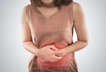
“肠系膜淋巴结炎”实际上就是指肠系膜淋巴结的炎性增生。多发生在右下腹,因为右下腹是回肠末段、盲肠和部分升结肠的系膜,也是淋巴系统最丰富的区域。
我们知道,肠道的感染性病变和一些全身疾病都会使得肠系膜淋巴结炎性增生,这一类的肠系膜淋巴结炎被称为继发性肠系膜淋巴结炎。
继发性淋巴结炎的原发疾病主要是各类肠道感染性病变,如:克罗恩病、回肠炎、结肠炎、憩室炎、阑尾炎等,系统性红斑狼疮等全身性疾病也可以导致继发性肠系膜淋巴结炎。
在被诊断为肠系膜淋巴结炎的患者中,利用 CT 检查,70%患者可以发现明显的肠道炎性病变,30%无明显的肠道炎性病变。而在有急腹症腹部症状的所有患者中 8.3%的患者可以发现右下腹肠系膜淋巴结肿大。由此可见,肠系膜淋巴结炎确实是急腹症的一个主要原因,但绝大部分都是继发性淋巴结炎,其基本的病变还是在肠道本身。
即使那 30%的“原发性”肠系膜淋巴结炎也需要进一步的研究。作者认为,这些病例都是轻度的感染性肠病继发的肠系膜淋巴结肿大,由于肠壁的炎性改变轻微常常被影像学首检医师忽视。欧美的研究者发现耶尔森肠炎菌是造成西方人末端回肠炎或结肠炎伴发肠系膜淋巴结炎的主要病原,而亚洲的研究者发现沙门氏菌似乎是东方人的主要病原。
有作者根据肠系膜淋巴结炎的自限性特征,认为肠系膜淋巴结炎可能是病毒感染,但到目前为止没有证据证实这点。
常见病因
- 便秘 — 便秘患儿可能表现为腹绞痛,有时这种疼痛可为重度。一项病例系列研究纳入 83 例儿童,这些儿童因急性腹痛而到初级保健机构或急诊科就诊,该研究表明急性或慢性便秘是腹痛最常见的基础病因,占 48%。在许多情况下,直肠检查是做出诊断的关键步骤。
- 具备以下特征中至少 2 项的儿童很可能为便秘:
- 每周大便<3 次
- 大便失禁(通常与功能神经性大便失禁有关)
- 直肠内或腹部检查时可触及大块粪便
- 憋便姿势(retentive posturing,也称为便潴留姿势),或排便疼痛感
- 胃肠道感染 — 急性胃肠炎患儿在开始腹泻前可能出现发热、重度腹部绞痛和弥漫性腹部压痛。无腹泻时,胃肠炎的诊断应视为排除性诊断。
- 小肠结肠炎耶尔森菌性胃肠炎 会出现局灶性右下腹痛和腹膜刺激征,这些表现可能在临床上不易与阑尾炎区分。
- 肠系膜淋巴结炎 — 肠系膜淋巴结炎为肠系膜淋巴结的炎性疾病,可表现为急性或慢性腹痛。因为肠系膜淋巴结通常位于右下腹,所以肠系膜淋巴结炎有时类似阑尾炎和肠套叠。一项病例系列研究纳入 70 例临床疑似急性阑尾炎的儿童,通过超声、临床病程观察或手术,有 16%的患儿最终诊断为肠系膜淋巴结炎。
- 肠系膜淋巴结炎的患病率可能在升高,这很可能与越来越多地使用诊断性影像学检查来评估腹痛患儿有关。肠系膜淋巴结炎是通过超声显示有腹部淋巴结(直径>8mm )而诊断。诊断性影像学示存在增大的淋巴结本身并不能排除阑尾炎的诊断;还有必要证实阑尾结构正常。
- 急性肠系膜淋巴结炎是自限性疾病。治疗采用支持治疗,包括疼痛治疗和充分补液。肠系膜淋巴结炎患儿出现的腹痛通常在 1-4 周内消退;但许多患儿的症状可最长持续 10 周。长时间存在症状的患儿,或者出现体重减轻或其他全身性症状的患儿,可能需要评估炎症性肠病、结核或恶性肿瘤。
- 卵巢囊肿破裂 — 类似阑尾炎或腹膜炎的急性重度腹痛可能是卵巢囊肿破裂所致。罕见情况下可发生危及生命的出血。
- 异物摄入 — 年幼儿童常会吞服较小、光滑的非食物性物体,一旦异物已通过幽门,通常容易被排出体外。吞服异物的儿童出现腹痛时,需要紧急评估肠梗阻或穿孔,特别是异物锐利(可能导致肠穿孔)、长度>5cm(可能造成肠梗阻)、为多枚磁体(可能造成两枚磁铁互相吸附时其间的一段肠壁嵌压)或为纽扣电池(可能释放腐蚀性物质)时。
- 腹绞痛 — 婴儿发生腹绞痛时可能表现为易激惹、哭闹或看起来为腹痛。
- 其他提示腹绞痛诊断的临床特征包括:
- 典型的阵发性哭闹,持续至少 3 周
- 哭闹通常在夜间
- 哭闹可随排气或排便而缓解
- 喂养正常
- 无相关症状
- 体格检查正常

今天我们来说说孩子肚子痛——肠系膜淋巴结炎的问题。
在临床中,郑医生经常遇到肚子痛的孩子,但不是急性疼痛,而是偶尔痛一下,然后过一段时间又不痛了,特别是在饭后反复发作。家长带孩子到医院检查就发现有肠系膜淋巴结炎。
其实,肠系膜淋巴结炎在中医来说是积滞引起的腹痛,从饮食上调整才是治疗的根本,药物只是辅助作用。
- 你会发现:很多这类患儿都有过度喂养的习惯,没办法消化就积滞在中焦,阻滞中焦气机,不通则痛。
- 所以我们要给孩子清淡饮食,不可再大鱼大肉,让中焦气机通畅了之后再慢慢添加。
然后每天帮孩子顺时针摩擦腹部,以孩子感觉温热为度,还可以加用一些消食健脾的食物,调理一段时间后就会慢慢好转了。

三岁的小美女楠楠最近老喊肚子疼,妈妈带她来看医生,检查后大夫说是肠系膜淋巴结炎;晚上九点多了五岁的硕硕肚子疼得直不起腰来,检查后诊断也是肠系膜淋巴结炎,立即给他做上了理疗,效果立竿见影,二十分钟后硕硕有了笑模样……最近很多类似的病例,我们就来给大家说一说,肠系膜淋巴结炎到底是个啥?
概念:是指由于上呼吸道感染引起的回肠及结直肠区急性肠系膜淋巴结炎。
病因:病毒性感染,主要由 Coxsackie B 病毒或其他病毒所引起。好发于冬春季。
表现:常见于 7 岁以下的儿童,在上呼吸道感染后,有咽痛,倦怠不适,继之腹痛,恶心、呕吐,发热,腹痛以脐周及右下腹多见,呈阵发性发作,有压痛和反跳痛,痛点不固定。
最重要的是,肠系膜淋巴结炎引起的腹痛,需要和阑尾炎、肠套叠、肠炎等鉴别一下,所以,有了以上症状要带宝宝看医生哦,最常做的辅助检查就是 B 超。
- 缓解疼痛症状,腹痛厉害的宝宝可以做做物理治疗,我们急诊科有激光、理疗,3-5 次即可。
- 宝宝在日常生活中要饮食规律,清淡,营养均衡,忌食辛辣刺激食物。不要暴饮暴食,避免吃过硬、过热、过冷、过分粗糙的食物。可以吃米饭、水果,多吃含纤维素的食物,少吃易产气的食物。
- 养成定时排便的习惯。
- 患病期间忌食冷饮、冰淇淋、咖啡、巧克力。
- 可以吃的水果、蔬菜:苹果、香蕉、菠菜、油菜、白菜等。
一般情况下,一周就能好啦。但肠系膜淋巴结炎容易反复,不严重的话,可以自行缓解,如果严重腹痛,伴有发烧、呕吐等,需要去医院看医生排除急腹症。

很多小孩子,可能都会经历这种疼痛。
它入侵身体的时候。有的小孩会突然大喊大叫,说肚子很痛,然后要蜷缩起来或者蹲着。有的一会说妈妈我肚子疼,然后呢你看他玩得又很嗨,但是过一会他又喊肚子疼了。并且这个疼痛是隐隐的,一般都是吃饭后或者运动过后会发作加重。
遇到这种情况,有的家长会给孩子喝点热水、或者加一点衣服,但也只能缓解一时甚至没效果。也有的家长,会给孩子吃“胃舒平”一类的药,但是吃了后效果也是不太好的,因为过不了多久又发作了。
还有的家长以为孩子是不想上幼儿园说肚子痛,因为过一会儿又不疼了,不好判断,也就没太引起注意。久而久之,孩子可能会出现低热、呕吐、腹泻或手脚冰凉的情况,有时也会有大便干,甚至羊屎蛋的情况。
其实出现这些情况,大多是因为没有弄清楚孩子腹痛的原因,看着孩子遭受疼痛的煎熬,妈妈们就会很焦虑。所以,我们需要了解清楚。
西医叫它
肠系膜淋巴结炎
西医把这种折磨小孩子的疼痛叫作肠系膜淋巴结炎。并认为,一般是病毒感染导致的。并且,通常是小孩子感冒后会出现。疼痛的部位,主要是在肚脐左右两侧偏下一点。
治疗方式,多是保守治疗和对症处理。比如,有明显的感染症状,可能会主张使用一些抗生素或者一些抗病毒药物。
但是多数情况下,都认为,这个问题不太需要治疗,在家保守处理就可以了。
中医认为
寒邪入侵是外在原因
在中医看来,寒邪如果比较强盛,入侵孩子的身体以后,会直接进入中下焦的脾胃、大小肠这些肚子的部位,待在那里,最后就会造成这些地方的津血堵在这里动不了。
我们知道,如果津液和气血堵在这里了,正邪气一打架,就会产生疼痛。这就是中医说的不通则痛。
但要清楚,寒邪阻滞是外因,是诱发因素。
脾胃虚弱
才是内在原因
其实,孩子肠系膜淋巴结炎的根本原因是正气不足了,而正气不足是因为脾胃虚弱导致的。因为脾胃虚弱了,津血也就虚了,在表,是不能抵抗外邪;在里,又驱赶不走邪气。所以也是会表现出腹部疼痛的。
脾是后天生化之本,像土地一样承载着我们的人体,让我们能够健康地成长,它虚了,我们的正气就会不足,就容易遭受外邪的侵袭。这其实和前面说的寒邪入侵的病因是互相关联和影响的。
治疗时主要是
温中散寒和补阳气
小儿出现不明原因腹痛的时候,家长可以先带去医院检查,排除危急重症。对于这种长期反复疼痛隐隐不剧烈的肠系膜淋巴结炎,可以考虑用中医的方式来治本。
主要是采取温中散寒和补阳气、脾气的方式。这样入侵的寒邪可以被赶走,而虚弱的脾胃也能够得到滋养和恢复。
父母在家
可以给孩子做推拿
成就感和性价比:小儿推拿是我们父母可以日常在家里就操作的,调理肠系膜淋巴结炎也很有效。只要你学会了,每天抽出10几分钟,就会有效果,并且是终生都受用的,大大节省了时间和开支。
安全性:小儿推拿是自然绿色疗法,不像吃药那样,无毒副作用,只要你跟着老师操作,知道什么可以做什么不可以做,就非常安全。
运动性:推拿其实还是有益身心的锻炼方式。在学习小儿推拿的过程中,不仅有利于你和孩子的沟通交流,而且可以使你和孩子放松身心,充分感受彼此。

以下内容来源于新英格兰医学杂志。
Presentation of Case

Differential Diagnosis
Movement Disorders
Seizures
Functional Movement Disorder
Dyskinesia
Limb-Shaking TIAs
Clinical Impression and Initial Management

Clinical Diagnosis
Dr. Albert Y. Hung’s Diagnosis
Pathological Discussion

Pathological Diagnosis
Additional Management
Final Diagnosis

以下内容来源于新英格兰医学杂志。
Presentation of Case
Dr. Christine M. Parsons (Medicine): A 75-year-old woman was evaluated at this hospital because of arthritis, abdominal pain, edema, malaise, and fever.
Three weeks before the current admission, the patient noticed waxing and waning “throbbing” pain in the right upper abdomen, which she rated at 9 (on a scale of 0 to 10, with 10 indicating the most severe pain) at its maximal intensity. The pain was associated with nausea and fever with a temperature of up to 39.0°C. Pain worsened after food consumption and was relieved with acetaminophen. During the 3 weeks before the current admission, edema developed in both legs; it had started at the ankles and gradually progressed upward to the hips. When the edema began to affect her ambulation, she presented to the emergency department of this hospital.
A review of systems that was obtained from the patient and her family was notable for intermittent fever, abdominal bloating, anorexia, and fatigue that had progressed during the previous 3 weeks. The patient reported new orthopnea and nonproductive cough. Approximately 4 weeks earlier, she had had diarrhea for several days. During the 6 weeks before the current admission, the patient had lost 9 kg unintentionally; she also had had pain in the wrists and hands, 3 days of burning and dryness of the eyes, and diffuse myalgias. She had not had night sweats, dry mouth, jaw claudication, vision changes, urinary symptoms, or oral, nasal, or genital ulcers.
The patient’s medical history was notable for multiple myeloma (for which treatment with thalidomide and melphalan had been initiated 2 years earlier and was stopped approximately 1 year before the current admission); hypothyroidism; chikungunya virus infection (diagnosed 7 years earlier); seropositive erosive rheumatoid arthritis affecting the hands, wrists, elbows, and shoulders (diagnosed 3 years earlier); vitiligo; and osteoarthritis of the right hip, for which she had undergone arthroplasty. Evidence of gastritis was reportedly seen on endoscopy that had been performed 6 months earlier. Medications included daily treatment with levothyroxine and acetaminophen and pipazethate hydrochloride as needed for cough. The patient consumed chamomile and horsetail herbal teas. She had no known allergies to medications, but she had been advised not to take nonsteroidal antiinflammatory drugs after her diagnosis of multiple myeloma.
Approximately 5 months before the current admission, the patient had emigrated from Central America. She lived with her daughter and grandchildren in an urban area of New England. She had previously worked in health care. She had no history of alcohol, tobacco, or other substance use. There was no family history of cancer or autoimmune, renal, gastrointestinal, pulmonary, or cardiac disease.
On examination, the temporal temperature was 37.1°C, the heart rate 106 beats per minute, the blood pressure 152/67 mm Hg, and the oxygen saturation 100% while the patient was breathing ambient air. She had a frail appearance and bitemporal cachexia. The weight was 41 kg and the body-mass index (the weight in kilograms divided by the square of the height in meters) 15.2. Her dentition was poor; most of the teeth were missing, caries were present in the remaining teeth, and the mucous membranes were dry. She had abdominal tenderness on the right side and mild abdominal distention, without organomegaly or guarding. Bilateral axillary lymphadenopathy was palpable. Infrequent inspiratory wheezing was noted.
The patient had swan-neck deformity, boutonnière deformity, ulnar deviation, and distal hyperextensibility of the thumbs (Fig. 1). Subcutaneous nodules were observed on the proximal interphalangeal joints of the second and third fingers of the right hand and on the proximal interphalangeal joint of the fourth finger of the left hand. Synovial thickening of the metacarpophalangeal joints of the second fingers was noted. There was mild swelling and tenderness of the wrists. She had pain with flexion of the shoulders and right hip, and there was subtle swelling of the shoulders and right knee. Pitting edema (3+) and vitiligo were noted on the legs. No sclerodactyly, digital pitting, telangiectasias, appreciable calcinosis, nodules, nail changes (including pitting), or tophi were present. The remainder of the examination was normal.

The blood levels of glucose, alanine aminotransferase, aspartate aminotransferase, bilirubin, globulin, lactate, lipase, magnesium, and phosphorus were normal, as were the prothrombin time and international normalized ratio; other laboratory test results are shown in Table 1. Urinalysis showed 3+ protein and 3+ blood, and microscopic examination of the sediment revealed 5 to 10 red cells per high-power field and granular casts. Urine and blood were obtained for culture. An electrocardiogram met (at a borderline level) the voltage criteria for left ventricular hypertrophy.

Dr. Rene Balza Romero: Computed tomography (CT) of the chest, abdomen, and pelvis, performed after the intravenous administration of contrast material, revealed scattered subcentimeter pulmonary nodules (including clusters in the right middle lobe and patchy and ground-glass opacities in the left upper lobe), trace pleural effusion in the left lung, coronary and valvular calcifications, and trace pericardial effusion, ascites, and anasarca. The scans also showed slight enlargement of the axillary lymph nodes (up to 11 mm in the short axis) bilaterally and a chronic-appearing compression fracture involving the T12 vertebral body.
Dr. Parsons: Morphine and lactated Ringer’s solution were administered intravenously. On the second day in the emergency department (also referred to as hospital day 2), the blood levels of haptoglobin, folate, and vitamin B12 were normal; other laboratory test results are shown in Table 1. A rapid antigen test for malaria was positive. Wright–Giemsa staining of thick and thin peripheral-blood smears was negative for parasites; the smears also showed Döhle bodies and basophilic stippling. Antigliadin antibodies and anti–tissue transglutaminase antibodies were not detected. Tests for hepatitis A IgG and hepatitis C antibodies were positive. Tests for hepatitis B core and surface antibodies were negative. A test for human immunodeficiency virus type 1 (HIV-1) and type 2 (HIV-2) was negative.
Findings on abdominal ultrasound imaging performed on the second day (Fig. 2A and 2B) were notable for a small volume of ascites and kidneys with echogenic parenchyma. Ultrasonography of the legs showed no deep venous thrombosis. An echocardiogram showed normal ventricular size and function, aortic sclerosis with mild aortic insufficiency, moderate tricuspid regurgitation, a right ventricular systolic pressure of 39 mm Hg, and a small circumferential pericardial effusion. Intravenous hydromorphone was administered, and the patient was admitted to the hospital.

On the third day (also referred to as hospital day 3), nucleic acid testing for cytomegalovirus, Epstein–Barr virus, and hepatitis C virus was negative, and a stool antigen test for Helicobacter pylori was negative. An interferon-γ release assay for Mycobacterium tuberculosis was also negative. Oral acetaminophen and ivermectin and intravenous hydromorphone and furosemide were administered.
Dr. Balza Romero: Radiographs of the hands (Fig. 2C through 2F) showed joint-space narrowing of both radiocarpal joints and proximal interphalangeal erosions involving both hands. Radiographs of the shoulders showed arthritis of the glenohumeral joint and alignment suggestive of a tear of the right rotator cuff. A radiograph of the pelvis showed diffuse joint-space narrowing of the left hip, without osteophytosis, and an intact right hip prosthesis.
Dr. Parsons: Diagnostic tests were performed, and management decisions were made.
Differential Diagnosis
Cancer
Infectious Disease
Autoimmune Disease
Hypocomplementemia
Dr. Beth L. Jonas’s Diagnosis
Pathological Discussion
Pathological Diagnosis
Discussion of Management
Follow-up
Final Diagnosis
Overlap syndrome of rheumatoid arthritis and systemic lupus erythematosus complicated by proliferative lupus nephritis, superimposed on amyloid A amyloidosis.

以下内容来源于PubMed。
Abstract
Sacituzumab govitecan (SG) significantly improved progression-free survival (PFS) and overall survival (OS) versus chemotherapy in hormone receptor-positive human epidermal growth factor receptor 2-negative (HR+HER2-) metastatic breast cancer (mBC) in the global TROPiCS-02 study. TROPiCS-02 enrolled few Asian patients. Here we report results of SG in Asian patients with HR+HER2- mBC from the EVER-132-002 study. Patients were randomized to SG (n = 166) or chemotherapy (n = 165). The primary endpoint was met: PFS was improved with SG versus chemotherapy (hazard ratio of 0.67, 95% confidence interval 0.52-0.87; P = 0.0028; median 4.3 versus 4.2 months). OS also improved with SG versus chemotherapy (hazard ratio of 0.64, 95% confidence interval 0.47-0.88; P = 0.0061; median 21.0 versus 15.3 months). The most common grade ≥3 treatment-emergent adverse events were neutropenia, leukopenia and anemia. SG demonstrated significant and clinically meaningful improvement in PFS and OS versus chemotherapy, with a manageable safety profile consistent with prior studies. SG represents a promising treatment option for Asian patients with HR+HER2- mBC (ClinicalTrials.gov identifier no. NCT04639986 ).
展开更多
